
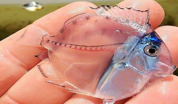
Transparent Juvenile Surgeonfish
Over the past several months, a photograph of a so-called "micro tang" has gone viral on more than one occasion, and the creature's strange appearance has sparked all sorts of questions from online commenters. Is the fish a fake? How big will this "micro" fish get? Is it dead? Is it a mutant?

First off, this isn't a "micro" anything – but it is a real fish. What you're looking at is a larval (baby) surgeonfish in the genus Acanthurus. In a matter of months, tiny wonders like this one grow up to be a respectable ten to 12 inches (about 30cm) long.

It seems this case of miniature mistaken identity can be traced to the Mr Good Travel Instagram page, who identified the fish as a "micro tang" in a post that racked up over 58,000 likes earlier this year. Before long, the fishy ID had surfaced on Reddit, Facebook and countless Pinterest boards.
The original image, however, belongs to Kevin Mattson, who notes that he released the fish alive, and in seemingly good condition, after the photo was taken.
Synopsis
- Yellowfin surgeonfish (Acanthurus xanthopterus) larvae were raised from wild-spawned eggs at 77-79F on cultured copepods, rotifers, artemia and microparticulate diets.
- The larval duration was 64 days.
- First record of Yellowfin surgeonfish culture.
Background
Everything about Acanthurus xanthopterus (Pualu in Hawaiian), is impressive. It is the largest Acanthurus species, growing up to 26 inches. Its distribution stretches from the lower Gulf of California all the way around the globe to East Africa. It lives in various reef habitats, sand slopes and lagoons and can occur as deep as 90 meters. It often travels in big schools. It feeds on everything from diatoms, detritus, filamentous algae and hydroids to small fish and crustaceans. It grows very quickly and undergoes intense changes in color as it matures. It can quickly change its color pattern, depending on its behavioral mood. A. xanthopterus is a beautiful fish! Unfortunately, it fits much better into a public aquarium than most hobbyist tanks due its size.

Culture
A. xanthopterus was cultured from eggs collected in Oahu’s coastal water in July 2018. This surgeonfish species was raised in a 20-gallon tank together with tilefish larvae, jack larvae, chub larvae, sole larvae, mahi-mahi larvae, boxfish larvae and snapper larvae. Preflexion larvae were offered copepod nauplii and rotifers. Postflexion larvae were fed artemia and micropellets in addition to copepods and rotifers (rotifers were discontinued on day 30).
After 28 days, one surgeonfish larva remained. Over the next 40 days, this fish survived multiple photo sessions and became a juvenile! On day 70, it was moved from the larval tank to a 40-gallon aquarium that also contained juvenile wrasses, porcupinefish, jacks and dragonets. It fed on pellets, nori, frozen seafoods and the natural diatom growth in the aquarium. This yellowfin surgeonfish now resides at the Waikiki Aquarium.
Development
A. xanthopterus eggs measured 0.7 mm in diameter. The larvae hatched at 1.8 mm TL (total length). They began to feed three days after hatching at 2.4 mm TL. Flexion occurred at 18 dph (5.1 mm TL). At 38 dph (11.9 mm TL), the larval body was deep and triangular-shaped with a silvery gut and a long 1st dorsal and anal spine.
At 50 dph (18.4 mm TL), the body was disk-shaped. At 58 dph (9.9 mm TL), the caudal peduncle spine was visible – a feature of the acronurus stage in surgeonfishes. From here on, the A. xanthopterus larva rapidly transformed into a juvenile. At 64 dph (26.4 mm TL), larval body was extremely laterally compressed and oblong with brown pigment on the dorsal body and anal fin. The fish behaved like juvenile at this time. By 70 dph, it was fully colored.
Conclusion
A. xanthopterus larvae are more robust and grow faster than A. triostegus larvae, under similar rearing conditions. Large fish species tend to be easier to culture and have shorter larval durations than their smaller relatives so this may be expected. Nevertheless; it’s exciting to discover these differences within the same genus – especially one as large, diverse and ecologically important as the genus Acanthurus – for which so little aquaculture research exists.
Recommended Videos
 Rare Genetic Mutation Makes This ‘Chimera’ Kitten One Of The Cutest Accidents Ever60 views
 Epic Beach Fails622 views
-
Advertisements
 Nicobar Pigeon Is The Closest Living Relative Of The Dodo96 views
 “Ridiculous Humans Doing Ridiculous Things”: 32 Bizarre Moments Captured And Shared On The Internet1553 views
 Bicycle Swallowed by Tree in Washington State124 views
 The colorful and fragrant blooming of Muscari flowers.117 views
 ‘Ugly Design’ Instagram Is Full Of Pics To Make You Laugh And Cringe And Here’s 50 Of The Bes5070 views
 Hilarious Beach Fails (30 GİF)11176 views
You may also like
 Experiencing the Allure of Exotic Durians: A Fascinating Exploration into Uncommon Delights
  Fabergé Eggs: Charming Jewelled Eggs Worth Millions Of Dollars
  20 Of The Most Hilarious Photos Of Folks Who Have Realized That Today Is The Unluckiest Day Of Their Lives
  “When Humans Leave”: 39 Pics Of Abandoned Places That Nature Started To Take Back